Fig. 4.
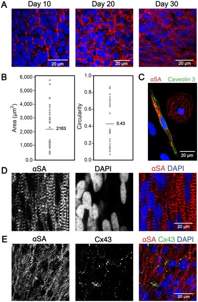
Ontogenetic tissue development generating well-defined and aligned sarcomeres over time. (A) Sarcomere definition and alignment became more pronounced with culture time. Immunofluorescence staining with cardiac marker αSA on days 10, 20, and 30 of differentiation showed increased sarcomere definition and alignment in 3D-dhECT CMs. (B) Day 52-60 dissociated 3D-dhECT CM area and circularity ranged from 400-5,800 μm2 (mean = 2,163 μm2) and 0.10 – 0.86 (mean = 0.43), respectively (n = 32). Blue highlighted data points indicate medians. (C) Representative day 52 dissociated CMs stained with αSA (red) and Caveolin 3 (green) for T-tubule development. Long-term cultured 3D-dhECT (day 124) CMs (D) developed highly aligned sarcomeres and contained large and elongated cell nuclei. (E) Additionally, these CMs expressed Cx43 on their transverse ends between adjoining cells.

