Figure 3.
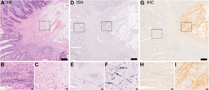
Human colorectal adenoma cells are devoid of decorin expression. Images of a representative human colorectal adenoma tissue sample. Panel consists of images of serial sections of the same tissue sample representing hematoxylin and eosin staining (A-C), in situ hybridization (ISH) (D-F) and immunohistochemistry (G-I) for decorin. Left frames in A, D and G mark areas of basal adenoma tissue and are magnified in B, E and H, respectively. Right frames in A, D and G mark stroma tissue and are magnified in C, F and I, respectively. In D and F, positive ISH signal for decorin can be seen in purple. Arrows in F indicate representative examples of positive ISH signals for decorin. In G and H, decorin immunoreactivity can be seen in brown color and localizes to the stroma of the tissue. Note that decorin mRNA and decorin immunoreactivity can only be seen in the stroma and they are totally absent from adenoma cells. Scale in A, D and G, 200 µm; in B, C, E, F, H, and I, 20 µm.

