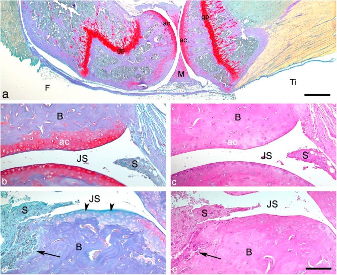
Figure 1.
The Bone-Inflammation-Cartilage (BIC) stain. (a) Histological section of a knee joint from a mouse with a case of mild septic arthritis, stained with the BIC stain. The proteoglycan content in the articular cartilage and growth plate stains red and the bone stains purple/blue. Scale, 500 μm. BIC- (b and d) and H&E (c and e)-stained sections of ankle joints from mice with septic arthritis. (b, c) Histology of a joint from a mouse with a mild case of arthritis. (d, e) Histology of a joint from a mouse with a severe case of arthritis, where synovitis, bone erosions and loss of articular cartilage are present. Note where the synovial inflammatory tissue invades the subchondral bone (arrows in d and e). The arrowheads in (d) indicate the loss of proteoglycan content in the articular cartilage (not visualized in H&E-stained section, d). Abbreviations: ac, articular cartilage; gp, growth plate; F, femur; M, meniscus; Ti, tibia; B, bone; JS, joint space; S, synovial membrane. Scacle, 100 μm.

