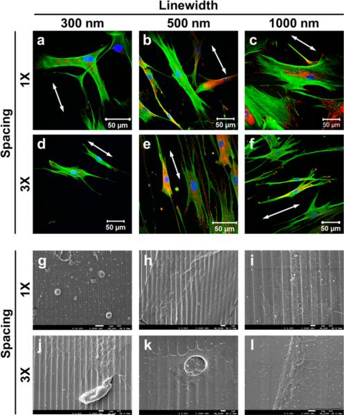
Figure 2.
Fibroblasts spreading on nanogratings of 150 nm in height. (a–f) Confocal images of the fibroblasts. The nuclei were stained with DAPI in blue, the actin filaments were stained with phalloidin in green, and focal adhesions were stained with paxillin in red. The white arrows point to the nanograting orientation. (g–l) SEM micrographs of the cell spread on nanogratings. The scale bars are 1 μm.

