
Figure 6. Model for coupling mechanism of InsP3R1 activation by InsP3.
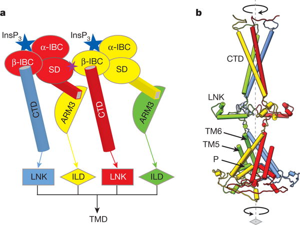
a, Schematic representation of inter-subunit contacts involved in propagation of InsP3-binding signal to the pore (colour-coded by subunit): from β-IBC to CTD/LNK domains and from the suppressor domain (SD) to ARM3/ILD domains of neighbouring subunits. b, The InsP3-induced changes in the LBDs can cause the helices in the cytosolic bundle to rearrange and trigger the motions of the LNKs, that may force the transmembrane bundle to adopt a conformation permeable for Ca2+; colour-coded by subunit.