Figure 2.

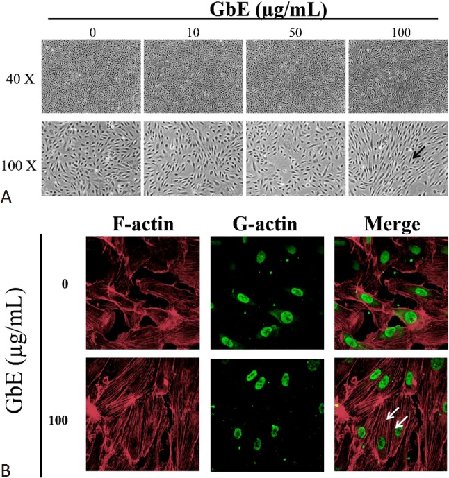
Ginkgo biloba extraction (GbE) altered cell morphology and cytoskeleton arrangement. Human umbilical endothelial cells (HUVECs) were treated with different concentrations of GbE or DMSO as control for 24 h. (A) Cell morphology gradually shifted from a typical cobble stone shape to a spindle shape (black arrow) when cells were incubated with increasing GbE concentrations. (B) Immunofluorescence Texas Red-X phalloidin staining for F-actin stress fibers revealed cytoskeletal rearrangement and increased stress fiber formation (white arrow) after treatment with 100 mg/mL of GbE. Alexa Fluor 488 DNase I conjugate was used for G actin staining to localize the nucleus. Images were acquired at 40X and 100X magnification. A confocal laser scanning microscope was used for the 400X magnification (n = 3).
