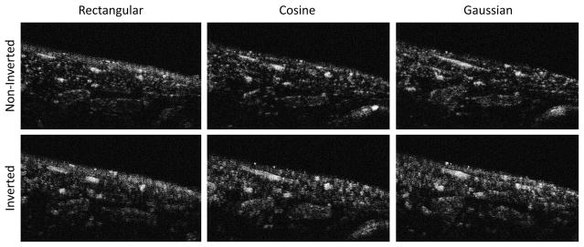
Figure 11.
Illustrative in vivo images after contrast enhancement formed by transmitting noninverted (top row) and inverted waveforms for rectangular, cosine, and Gaussian windowed pulses (left to right). The same slice of the same animal shown in Fig. 6A is displayed here for direct comparison. Results after contrast injection in all animals are summarized in Fig. 5B. While the image formed with the non-inverted Gaussian waveform performs best at rejecting tissue, the inverted cosine waveform appears to provide enhanced sensitivity at the cost of reduced resolution. The length of the scale bar is 4 mm.

