Figure 7.

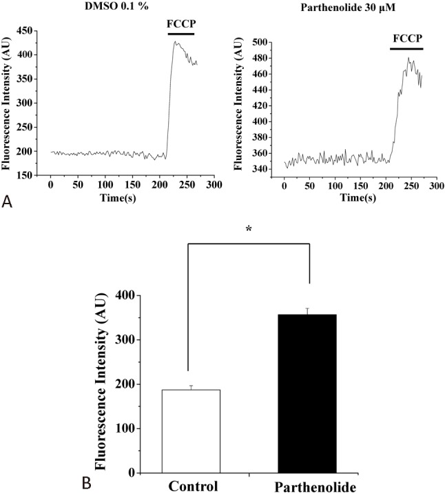
Parthenolide depolarized mitochondrial membrane potential in H9c2 cells. (A) Cells were pretreated with (right panel) or without (left panel) parthenolide (30 μM) for 2 h and then assayed for mitochondrial membrane potential using rhodamine 123 as dye. For authentication of the mitochondrial signals, FCCP (2 μM) was used to collapse mitochondrial membrane potential. (B) Quantification of results from (A). Results are mean ± standard error of the mean of 37-47 cells from 4 experiments. * indicates significant difference (p < 0.05) from the control. DMSO, dimethyl sulfoxide; FCCP, carbonyl cyanide4-trifluoromethoxyphenylhydrazone.
