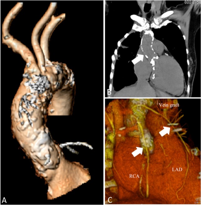
Figure 1.

A multi-slice computed tomographic scan shows diffuse atherosclerotic changes with heavy calcifications of the ascending thoracic aorta (A and B, arrow). The saphenous-vein grafts to left anterior descending artery (LAD) and right coronary artery (RCA) that had been placed during coronary artery bypass grafting three years earlier were patent (C, arrows).
