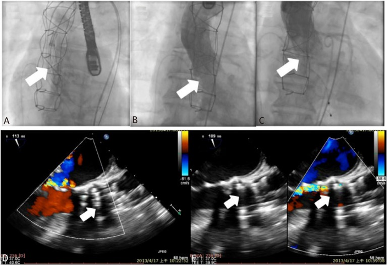
Figure 2.
Fluoroscopic images during the implantation of the CoreValve (A, arrow) and the immediate post-procedure aortogram showing severe aortic regurgitation (B, arrow). Intra-operative post-procedural transesophageal echocardiography demonstrated an under-expanded and mal-apposed CoreValve due to interference of the nodular calcification at the level of the aortic valve ring, leading to both severe valvular and paravalvular aortic regurgitation (D). After post-dilatation of the implanted CoreValve, both the immediate post-procedural aortogram (C, arrow) and transesophageal echocardiography (E, arrows) demonstrated a well-functioning CoreValve and mild aortic regurgitation.

