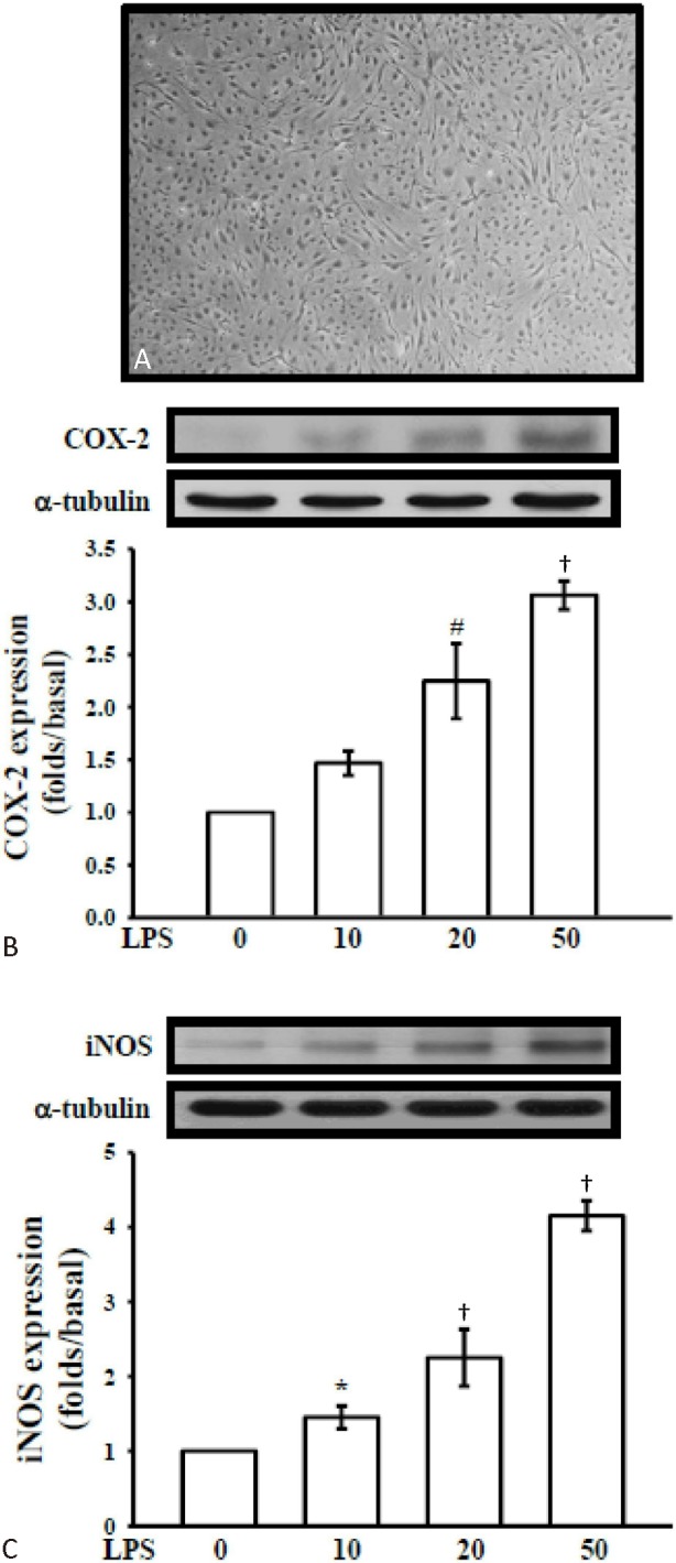
Figure 3.

LPS-induced expressions of cyclooxygenase-2 (COX-2) and inducible nitric oxide synthase (iNOS) in cerebral endothelial cell (CEC)s. (A) Morphology of primary cerebral endothelial cell. Cerebral endothelial cells (5 × 10⁵ cells in 6-well plates) were treated with vehicle (0.5% DMSO) or various concentrations of lipopolysaccharide (LPS) (10, 20, and 50 μg/ml) for 24 hr. Cell lysates were obtained and analyzed for (B) COX-2 and (C) iNOS protein expression by Western blotting. α-tubulin normalized to the resting condition. Data are shown as the mean ± SEM of three independent experiments.* p < 0.05, # p < 0.01 and † p < 0.001, compared with the resting group.
