
Figure 2.
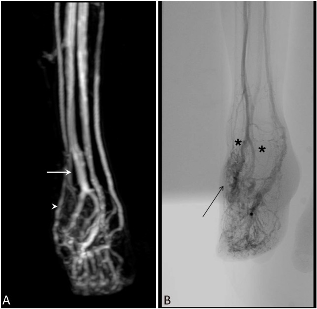
MRI revealed multiple arteriovenous communications between the dorsalis pedis artery (A, arrow) and superficial venous system (A, arrow head). Angiography showed extensive distribution of the arteriovenous malformation, with early engorged venous drainage, two major feeding arteries (B, astro) arising from the right pedis dorsalis artery, pedis plantaris artery, and its branches (B, arrow). MRI, magnetic resonance imaging.