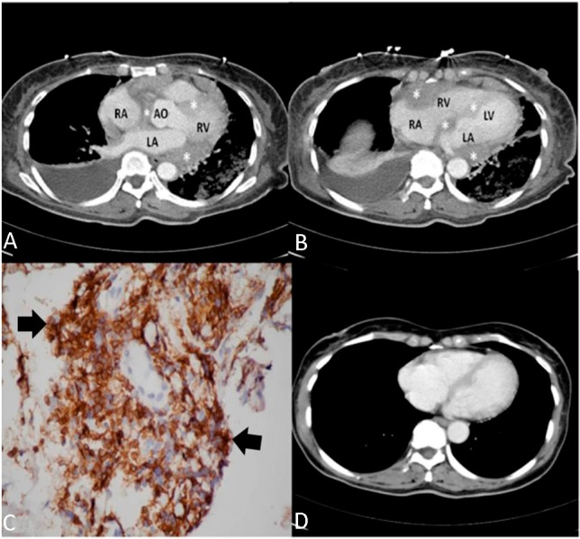
Figure 2.

Primary cardiac T-cell lymphoma. (A and B) An axial contrast-enhanced computed tomography (CT) scan revealed a diffuse, low-density mass (asterisks) involving the right ventricle, right and left atria, and atrioventricular septum, as well as compressing the aortic root. (C) An immunohistochemical stain (arrow, with brown and tan color) was positive for CD45RO. (D) A follow-up CT scan demonstrates regression of the mass after sixth months. AO, aorta; LA, left atrium; LV, left ventricle; RA, right atrium, RV, right ventricle.
