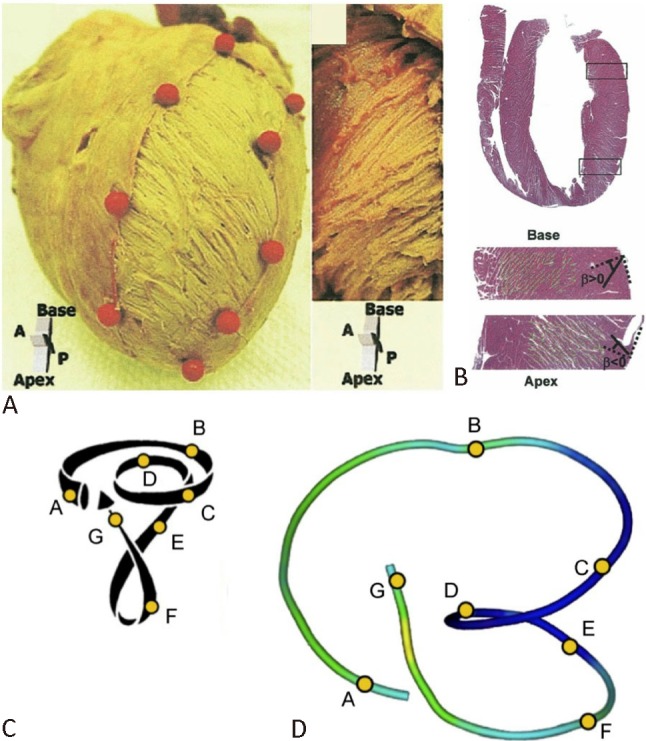
Figure 2.

Helical arrangement of myofibers in left ventricle. (A) Left-handed helix in the subepicardium and right-handed helix in the subendocardium in the left ventricle of an explanted porcine heart. (B) Longitudinal cross section of left ventricle fixed in diastole (hematoxylin and eosin stain) and radial orientation of the cleavage planes. (C) Torrent-Guasp helical ventricular myocardial band model. (D) Tractography reconstruction of diffusion tensor magnetic resonance imaging. Reproduced from Sengupta PP et al.8 and Poveda F et al.9 with permission.
