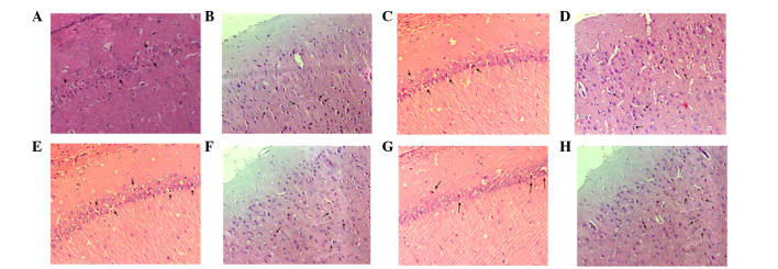
Figure 7.
Pathological examination of hematoxylin and eosin staining in the hippocampal CA1 and the temporal cortex 7 days after return of spontaneous circulation (magnification, ×200). In each recovery group there was strong nuclear staining, vacuolar changes associated with nerve damage, and eosinophilic cells (indicated with arrows). (A) CA1 and (B) temporal cortex in the control group, (C) CA1 and (D) temporal cortex in the intra-carotid administration group, (E) CA1 and (F) temporal cortex in the femoral venous infusion group, and (G) CA1 and (H) temporal cortex in the lateral ventricle administration group. CA1, region I of hippocampus proper.

