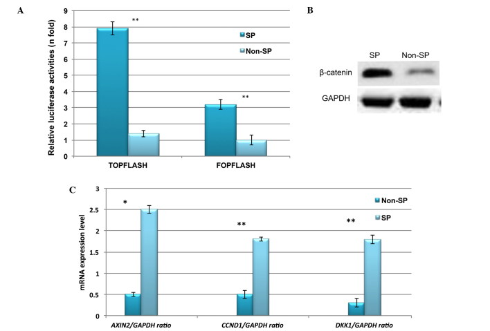
Figure 5.
Constitutive expression of Wnt/β-catenin signaling in liver cancer SP cells. (A) TOPflash and FOPflash assay indicating that β-catenin is markedly upregulated in liver cancer SP cells. (B) Western blot image indicating the increased β-catenin protein expression in SP cells. (C) Reverse transcription-quantitative polymerase chain reaction analysis indicating the increased mRNA expression of Wnt/β-catenin target genes in SP cells. SP, side-population; GAPDH, glyceraldehyde 3-phosphate dehydrogenase; AXIN2, axis inhibition protein 2; CCND1, cyclin D1; DKK1, dickkopf Wnt signaling pathway inhibitor 1. *P<0.05 and **P<0.01 vs. non-SP cells.

