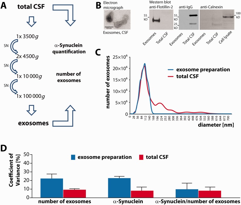
Figure 1.
Isolation and characterization of exosomes from CSF. (A) Exosomes were isolated by subsequent centrifugation rounds including a final 100 000g ultracentrifugation step from a CSF volume of 0.5 ml. Exosome numbers and α-synuclein content were quantified in total CSF and exosome fractions by nanoparticle tracking analysis and by electrochemoluminescence assay. (B) Left: Electron microscopy of the 100 000g exosome pellet derived from 4 ml of CSF. Scale bar = 100 nm. Right: For western blot analysis exosome pellets were prepared from 2.5 ml of CSF and resuspended in 20 µl of sample buffer. CSF was diluted 1:5 in sample buffer and 20 µl of the exosome preparation and 20 µl of total CSF were probed with an antibody against the exosomal marker protein flotillin-2 (left panel). As a negative control, we probed exosome preparations and CSF with a secondary antibody against human IgG (middle panel). To rule out microsomal contamination of the exosome preparation, 20 µl of the exosome pellet, total CSF and of a cell lysate of mouse neuroblastoma N2a cells were blotted and incubated with an antibody against the endoplasmatic reticulum protein calnexin (right panel). (C) Exosome numbers were determined by nanoparticle tracking analysis in both, total CSF and the 100 000g exosome pellet derived from 0.5 ml CSF after resuspension in PBS. A representative plot depicting vesicle size and number of vesicles is shown (total CSF: red curve; corresponding exosome pellet: blue curve; peak: 102 nm). The values were adjusted for the respective dilution factors and calculated to represent the absolute vesicle numbers in 1 ml of CSF and in exosomes derived from the same CSF volume. (D) The coefficient of variance was determined for the number of exosomes in the exosome preparation and in total CSF, for the amount of exosomal α-synuclein protein and total CSF α-synuclein protein, and for the ratio of exosomal α-synuclein protein levels to the number of exosomes. Blue bars: measurements in the exosome preparation; red bars: measurements in total CSF. CSF samples from two different patients were analysed in replicates of n = 3 and n = 4.

