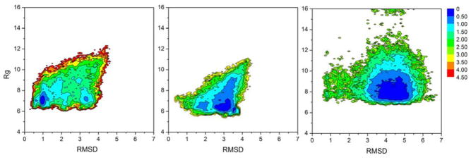
Figure 2.
Free energy landscapes of three fragments at 300K from REMD explicit water simulations. (A) Rg vs RMSD (backbone 43 – 49) to the native HP36 structure for HP-1 (B) Rg vs RMSD (backbone 54–59) to the native HP36 for HP-2 (C) Rg vs. RMSD (backbone 64–70) to the native HP36 for HP-3. While all three fragments remain compact as in HP36, only HP-1 has a free energy minimum located at a low RMSD. The other two fragments occupy minima with higher RMSDs and more broad minima than HP-1.

