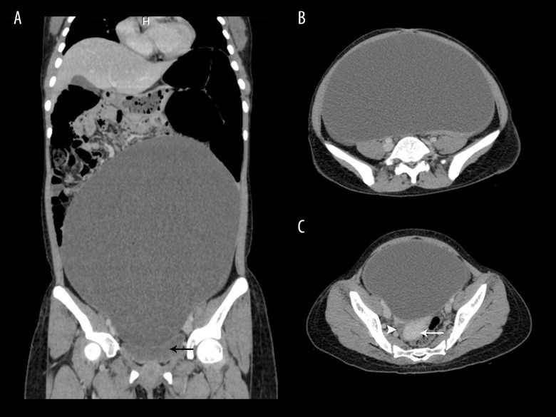
Figure 3.
Contrast-enhanced computed tomography of the abdomen and pelvis, in coronal reformatted (A) and axial (B, C) sections reveal a large, unilocular, purely cystic lesion arising from the pelvis and occupying almost the entire abdominal cavity. Note the absence of enhancing soft tissue or septae within the lesion. Also note the urinary bladder (black arrow), uterus (white arrow) and right ovary (white arrowhead). The left ovary could not be visualised separately from the lesion implying left ovarian origin of the cystic lesion.

