

Abstract
Aim:
The objective of this study is to investigate the hypoglycemic, hypolipidemic, and hepatoprotective effects of the aqueous extract of parsley, basil, and chicory whole plant in normal and dexamethasone (Dex) rats.
Materials and Methods:
50 female albino rats were used in this study and divided into 5 groups (for each 10). Group (1) fed basal diet and maintained as negative control group. Group (2) received Dex in a dose of (0.1 mg/kg b. wt.). Groups 3, 4, and 5 were treated with Dex along with three different plant extracts of parsley, basil, and chicory (2 g/kg b. wt.), (400 mg/kg b. wt.), and (100 mg/kg b. wt.), respectively.
Results:
All these groups were treated given three times per week for 8 consecutive weeks. Dex-induced alterations in the levels of serum glucose, triglyceride, cholesterol, low-density lipoprotein-cholesterol levels and cardiovascular indices and serum alanine aminotransferase, aspartate aminotransferase and lactate dehydrogenase activities, liver thiobarbituric acid (TBARS) levels increased, while high-density lipoprotein-cholesterol, total protein, albumin, and liver glutathione (GSH) levels decreased. On the other hand, plant extracts succeeded to modulate these observed abnormalities resulting from Dex as indicated by the reduction of glucose, cholesterol, TBARS, and the pronounced improvement of the investigated biochemical and antioxidant parameters.
Conclusions:
It was concluded that probably, due to its antioxidant property, parsley, basil, and chicory extracts have hepatoprotective effects in Dex-induced in rats.
KEY WORDS: Antioxidants, dexamethasone, hyperglycemia, hyperlipidemia
INTRODUCTION
Dexamethasone (Dex) has potent immunosuppressant and anti-inflammatory properties but therapeutic benefits of this drug limited as it presents several side effects such as insulin resistance and skeletal muscle atrophy and generation of the free radicals which may contribute to oxidative stress as long-term treatment [1]. Natural products have been examined for promising new source pharmaceutical and therapeutic agents in plant secondary metabolites that characterize definite plants [2].
It is thought that the health promoting effect of Petroselinum crispum (parsley) may be due to its flavones components as ascorbic acid, carotenoids, flavonoids, coumarins, myristicin, apiol, various terpenoid compounds, phenylpropanoids, phthalides, furanocoumarins, and tocopherol [3].
Ocimum basilicum (basil) had been found to contain linalool, eugenol, methyl chavicol, methyl cinnamate, ferulate, methyl eugenol, triterpenoids, and steroidal glycoside known to exhibit antioxidant, chemopreventive, anti-inflammatory, bactericidal, antiulcer activities, a nervous system stimulant effect, modulatory effect on glutathione and antioxidant enzymes, antidiarrheal and hypoglycemic effects [4,5].
Cichorium intybus L. (chicory) own excessive medicinal importance as it has alkaloids, inulin, sesquiterpene lactones, coumarins, vitamins, chlorophyll pigments, unsaturated sterols, flavonoids, saponins, and tannins [6].
The aim of this study was both to investigate the hepatoprotective effect of P. crispum, O. basilicum, and C. intybus L. aqueous extracts against Dex-induced in rats and to explore the antioxidant capability of the extracts.
MATERIALS AND METHODS
Chemicals
Dex ([Fortecortine® 8 mg - Mono ampule) Manufactured by Sigma - Tec Pharmaceutical industries - Egypt - S. A. E. under Licence of: Merck KGaA, Darmstadt, Germany).
Plant Materials
Parsley (P. crispum), basil (O. basilicum), and chicory (C. intybus L.) leaves were collected from herbal medicine market (Cairo, Egypt) and identified by an ecologist in plant department, Faculty of Science, Beni-suef University. The leaves of these plants were cleaned, shade dried for 30 days at room temperature, crushed to a coarse powder and preserved for further processing.
Preparation of Plant Extract
Preparation of aqueous parsley extract
The 100 gram dried parsley leaves were extracted by adding 1000 ml of distilled water and boiled for 30 min. The extract was then filtered, and then the filtrate was evaporated, using rotary evaporator under reduced pressure to dryness (at 45°C). The extract was dissolved in distilled water before the administration to rats [7].
Preparation of aqueous basil extract
The 300 g ground powder of dried basil leaves was infused for 30 min in 200 ml of distilled water at 100°C followed by filtration. The solution obtained was concentrated rotary evaporator under a vacuum at 65°C. The resulting crude extract was suspended in 30 ml sterile distilled water, and aliquots were stored at −20°C until use [8].
Preparation of aqueous chicory extract
The powdered chicory leaves were added to the already boiling distilled water and infused for 15 min. Then, the infusion (2% w/v) was filtered, and the filtrate was freshly used [9].
Animals
Female albino rats (Rattus norvegicus) weighing about 120-150 g were used for the study and were kept in the animal house at 26 ± 2°C with relative humidity 44% to 56% along with light and dark cycles of 12 h. Animals were provided with standard diet and water ad libitum.
Animal Groups
For the achievement of the objectives of this study, 50 female albino rats were randomly divided into 5 groups including the normal groups and the diabetic groups each group consists of 10 rats. Group 1: Served as normal control. Group 2 was considered as Dex-control was given Dex subcutaneously 0.1 mg/kg/day [10], and this group was considered as control for the Groups 3, 4, and 5. Group 3 (Dex-treated with parsley): The rats in this group were given Dex and also were administrated aqueous parsley at dose level of 2 g/kg/b. wt. [7]. Group 4 (Dex-treated with basil): The animals in this group were Dex and also were given aqueous basil at dose level 400 mg/kg/b. wt. [4]. Group 5 (Dex-treated with chicory): The animals in this group were Dex and also were treated with aqueous chicory at dose level 100 mg/kg/b. wt. [11]. All these groups were treated for three times per week for 8 consecutive weeks and the treatments with parsley, basil, and chicory were performed orally between 7.00 and 9.00 a.m.
Biochemical Assay
At the end of the experimental period (8 weeks), rats were sacrificed under diethyl ether anesthesia. Blood samples were collected from each rat, allowed to coagulate at room temperature then centrifuged at 3000 r.p.m. for 20 min. The clear, non-hemolyzed supernatant sera were quickly removed and kept at −20°C until examined.
The serum samples were analyzed for glucose according to the method of [12] using standard diagnostic kits from Spinreact and serum cholesterol [13], triglycerides [14], high-density lipoprotein-cholesterol (HDL-C) [15], total protein [16], albumin [17] using the commercial assay kit provided from Bioscope Diagnostics, beta lab, Egypt, while very low-density lipoprotein-cholesterol (VLDL-C), LDL-C, and cardiovascular indices were calculated mathematically according to [18,19] using the following formula:
VLDL-C = Triglycerides/5
LDL-C = Cholesterol - (Triglyceride/5) - HDL-C
Cardiovascular index 1 = Total cholesterol/HDL-C.
Cardiovascular index 2 = LDL-cholesterol/HDL-C.
The activity of alanine aminotransferase (ALT) and aspartate aminotransferase (AST) was determined kinetic at 546 nm using a standard method [20], respectively, using kit provided from Bioscope Diagnostics, betalab, Egypt. Lactate dehydrogenase (LDH) was measured using the kinetic method described by Young [21] using reagent kits obtained from BioSystems, Barcelona, Spain.
Hepatic Oxidative Stress and Antioxidant Enzymes Assay
Liver tissues were homogenized in cold 0.9% NaCl with a glass homogenizer to make up to 10% homogenate (w/v). The homogenates were centrifuged, and the clear supernatants were used for lipid peroxidation (LPO), glutathione (GSH), and antioxidant enzymes. The supernatant was used for the estimation of malondialdehyde (MDA) [22]; GSH [23]; GSH s-transferase (GST) [24]; GSH peroxidase (GPx) [25]; GSH reductase (GR) [26]; and catalase [27] levels using the reagent kits purchased from Biodiagnostic Company, Giza, Egypt.
Histological Examination of Liver
Liver specimens were fixed in 10% formal saline for 24 h. 5% formic acid was used for decalcification, processed, and embedded in paraffin. Thin paraffin sections (4 µm) were stained by H and E [28].
Statistical Analysis
The data were analyzed using the one-way analysis of variance [29] followed by least significant difference test to compare various groups with each other. Results were expressed as mean ± standard deviation (SD), and values of P > 0.05 were considered non-significantly different, while those of P < 0.05, P < 0.01, and P < 0.001 were considered significantly, highly, and very highly significantly different, respectively.
RESULTS
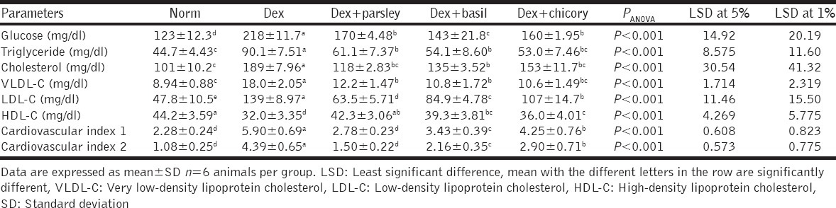
Data in Table 1 show treatment of diabetic rats induced by Dex with parsley, basil, and chicory extracts caused considerable positive effects on the investigated parameters as compared to their corresponding control rats where P < 0.001 in all compared groups.
Table 1.
Serum biochemical parameters in control and different treated groups
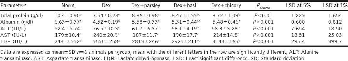
Data presented in Table 2 demonstrated that treatment of diabetic rats induced by Dex with parsley and chicory extracts cause a significant increase in serum total protein and albumin while treatment with basil cause a non-significant increase in total protein and a significant increase in albumin level when compared with diabetic rats.
Table 2.
Serum liver function markers in control and different treated groups
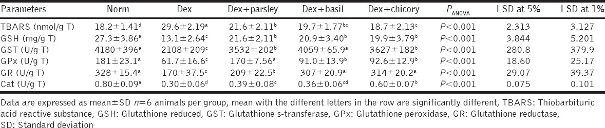
Results showed that the treatment of rats with parsley, basil, and chicory extracts produced a profound, significant decrease of the LPO products in the liver and also, produced an increase of glutathione level and antioxidant enzymes when compared to their control group as shown in Table 3.
Table 3.
Liver oxidative stress marker and antioxidant parameters in control and different treated groups
Histopathological Results
Microscopically, liver of rat from negative control revealed no histopathology change [Figure 1a]. Meanwhile, section of liver of rat from positive control group (rats administrated with Dex) revealed fatty change was detected in diffuse manner all over the hepatocytes in association with inflammatory cells infiltration in the portal area [Figure 1b]; a section of liver of parsley extract treated rats showed there was no histopathological alteration [Figures 1c and d]: The liver of rats from group supplemented with aqueous basil extract showed normal liver with few individual hepatocytes [Figure 1e]. Moreover, the liver of rats from group supplemented aqueous chicory revealed no histopathological changes except mild congestion was observed in the central veins [Figure 1f].
Figure 1.
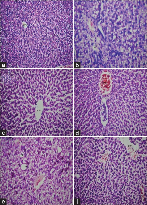
(a-f) Photomicrographs of rat liver sections of different experimental groups stained with hematoxylin and eosin, (a and b) liver sections of normal (G1) and dexamethasone (G2) groups, (c-d) liver sections of diabetic rats treated with parsley extract group (G3), (e) Liver section of diabetic rats treated with basil extract group (G4), and (f) liver section of diabetic rats treated with chicory extract group (G5)
DISCUSSION
The causes for diabetes after chronic Dex-treatment explained by reduced insulin sensitivity, impaired α and β-cell functions, increased hepatic gluconeogenesis, by stimulating amino acids releasing from skeletal muscle, fatty acids and glycerol from adipose tissue and enhance gluconeogenic enzymes expression as phosphoenolpyruvate carboxykinase [30].
Hyperlipidemia caused in Dex-induced diabetes is due to excess mobilization of fat from the adipose tissue, increase VLDL secretions by the liver and stimulate VLDL formation by the intestine, decreasing hepatic lipoprotein lipase activity that inhibit the removal of plasma VLDL and leading to an increase plasma VLDL level [31].
A decline in serum total protein level in diabetics due to decrease in protein synthesis, increase in catabolic processes, and reduction of protein absorption as inhibition of oxidative phosphorylation [32].
Corticosteroids therapy associated with several forms of liver injury leads to increase the liver function enzymes. ALT only significantly elevated in hepatobiliary disease and is liver specific enzyme AST level increased with damages of heart or skeletal muscle as well as of liver parenchyma. Changes in LDH may be attributed to severe damage to heart tissue as myocardial necrosis [33-35].
The Dex-induced oxidative stress and altered the antioxidant status in several tissues. The reduction of GSH contents corresponds to cellular oxidative damage and death as the cellular energy is impaired or due to the fast oxidation of GSH-induced by steroidal drug Dex. Depletion in the activity of antioxidant enzymes can be due to an enhanced radical production during Dex metabolism [36,37].
The current biochemical alterations were coincided with the present histological investigation for liver section, so the negative results associated with Dex may be attributed to the hepatic injury induced by superoxide anions and hydroxyl radicals which cause oxidative damage to cell membrane resulted in fatty change in diffuse manner all over the hepatocytes in association with inflammatory cells infiltration in the portal area. In agreement with our result [38] showed the treatment of rats with glucocorticoids led to the accumulation of lipids in the liver.
Parsley extract possess antihyperglycemic by facilitating glucose usage via extra-pancreatic ways as inhibition of gluconeogenesis and may be stimulate glycolysis process directly [6]. Aqueous extract of parsley possesses hypocholesterolemic and hypotriglyceridemic activities due to it has flavonoids that possess the bioactivity to beneficially affect the cardiovascular risk factors such as lipoprotein oxidation, dyslipidemia, endothelial dysfunction, and blood platelet aggregation and regulation of lipid levels in plasma or tissue leads to a decrease in the risk of micro or macro vascular disease and related complications [39]. Flavonoids decreased blood cholesterol levels by decreasing the biosynthesis of cholesterol, enhancing the phosphorylation of 3-hydroxy-3-methylglutaryl coenzyme A reductase indirectly [40].
Phytochemical screening of parsley has revealed the presence of flavonoids (apiin, luteolin, and apigenin-glycosides) and a considerable elevation in albumin concentration was recorded in apigenin-treated mammary adenocarcinoma group [41]. The parsley showed a significant decrease in the serum activity of AST, ALT, and LDH this result indicated that parsley had able to regenerate liver after liver cell damage and reduced incidence of heart disease in diabetes mellitus as it contains flavonoids, particularly the quercetin (as a flavonol) [42]. A variety of flavonoids, lignans, alkaloid, bisbenzyl, coumarins, and terpenes were tested for their antioxidant activity as showed they able to prevent the oxidative damage in epithelial cells kidney and liver [43]. Parsley extract has the ability to reduce the toxic effects may be recognized to the high nutritive value that determined too high percent of vitamins (A, C, riboflavin, and niacin) and minerals (Fe, Mg, P, K, Ca, Na, and Zn) [44].
O. basilicum decreased glucose levels in blood which act via inhibition renal glucose reabsorption and/or inhibition of hepatic glucose production, improving insulin action or stimulate peripheral tissues to utilize glucose [45]. The reduction of plasma total cholesterol and its LDL fraction by the O. basilicum extract was associated with an increase in plasma HDL-cholesterol, and the activity of O. basilicum extract to decrease cholesterol concentration by stimulating the elimination of cholesterol in the form of bile acids by promoting cholesterol mobilization from peripheral tissues to the liver [46]. There is a significant increase in the amount of protein and globulin levels as O. basilicum is immunostimulant herbals incorporated diets helped to increase the humoral elements in the serum [47]. Basil extract decreases the activities of serum AST and ALT due to it increase the level of antioxidant enzymes that may protect liver against the damaging effects and inhibit LPO [48]. Basil is a rich source of flavonoids, and the hepatoprotective effect of O. basilicum may be attributed to the antioxidant activity of its flavonoids [4], so the liver of rats from the group treated with aqueous basil extract showed normal liver.
Water-soluble extract of chicory reduced serum glucose may be due to the presence of inulin, oligofructose, and esculetin [49,50]. The decrease in blood cholesterol level might be due to chicory has the ability to stimulate lactic acid producing bacteria which secret the hydrolase enzyme that in-turn converts bile salts into deconjugated bile acids and ultimately resulted in the reduced serum cholesterol level, and the insulin decrease the expressions of acetyl-coenzyme A carboxylase and fatty acid synthase messenger RNA [49,51]. Inulin and oligofructose decrease the synthesis of triglycerides and fatty acids in the liver and decreasing their level in serum by inhibiting hepatic lipogenesis and reducing the risk of atherosclerosis [34]. Significant improvement in albumin serum level of chicory protected rats compared to nitrosamine precursors-treated rats [52]. The administration of chicory supplemented diet resulted in an improvement of protein pattern by preventing protein oxidation and improves liver and other organs functions which synthesized plasma protein [34,50]. The antihepatotoxic effect of chicory due to it contains isoflavones, polyphenols, and other antioxidants that can reduce the activity of serum ALT and AST, and it was noticed that chicory significantly lowers serum activity of AST and ALT enzymes in carbon tetrachloride (CCL4) intoxicated rats. Liver function was improved due to protective activity of antioxidants components in chicory extract, and it was shown that chicory decreased levels of MDA and increased GSH, antioxidant enzymes in (CCL4) intoxicated rats [52-55]. Chicory cause elevation of intracellular antioxidant enzyme activities and decreased oxidative stress in tissues due to chicory extract enhance endogenous antioxidant defense status [49,56]. This hepatoprotection of chicory prevent liver damage, and no histopathological changes occur.
CONCLUSION
The parsley, basil, and chicory extracts offered a hepatic protection against Dex-induced in rats. In a comparison of our result, we found that parsley has a potent hepatoprotective effect more than basil and chicory. All the aforementioned effects of extracts may explain their ameliorative impact on Dex changes in our study.
ACKNOWLEDGMENTS
The authors are thankful to the faculty of Science, Beni-Suef University for help in conducting this study and providing all required facilities. The authors are grateful to Professor Dr. Adel Mohamed Bakeer Kholoussy, Professor of Pathology, Faculty of Veterinary Medicine, Cairo University, for reading the histopathological sections.
Footnotes
Source of Support: Nil
Conflict of Interest: None declared.
REFERENCES
- 1.Zanchi NE, Guimarães-Ferreira L, de Siqueira-Filho MA, Felitti V, Nicastro H, Bueno C, et al. Dose and latency effects of leucine supplementation in modulating glucose homeostasis: Opposite effects in healthy and glucocorticoid-induced insulin-resistance states. Nutrients. 2012;4:1851–67. doi: 10.3390/nu4121851. [DOI] [PMC free article] [PubMed] [Google Scholar]
- 2.Saha S, Mukhopadhyay MK, Ghosh PD, Nath D. Effect of Methanolic Leaf Extract of Ocimum basilicum L. on benzene-induced hematotoxicity in mice. Evid Based Complement Alternat Med. 2012;2012:176385. doi: 10.1155/2012/176385. [DOI] [PMC free article] [PubMed] [Google Scholar]
- 3.Petrolini FV, Lucarini R, de Souza MG, Pires RH, Cunha WR, Martins CH. Evaluation of the antibacterial potential of Petroselinum crispum and Rosmarinus officinalis against bacteria that cause urinary tract infections. Braz J Microbiol. 2013;44:829–34. doi: 10.1590/S1517-83822013005000061. [DOI] [PMC free article] [PubMed] [Google Scholar]
- 4.Gbadegesin MA, Odunola OA. Aqueous and ethanolic leaf extracts of Ocimum basilicum (sweet basil) protect against sodium arsenite-induced hepatotoxicity in Wistar rats. Niger J Physiol Sci. 2010;25:29–36. [PubMed] [Google Scholar]
- 5.Dasgupta T, Rao AR, Yadava PK. Chemomodulatory efficacy of basil leaf (Ocimum basilicum) on drug metabolizing and antioxidant enzymes, and on carcinogen-induced skin and fore stomach papilloma genesis. Phytomed. 2004;11:139–51. doi: 10.1078/0944-7113-00289. [DOI] [PubMed] [Google Scholar]
- 6.Abbas ZK, Saggu S, Sakeran MI, Zidan N, Rehman H, Ansari AA. Phytochemical, antioxidant and mineral composition of hydroalcoholic extract of chicory (Cichorium intybus L.). leaves. Saudi J Biol Sci. 2015;22:322–6. doi: 10.1016/j.sjbs.2014.11.015. [DOI] [PMC free article] [PubMed] [Google Scholar]
- 7.Ozsoy-Sacan O, Yanardag R, Orak H, Ozgey Y, Yarat A, Tunali T. Effects of parsley (Petroselinum crispum) extract versus glibornuride on the liver of streptozotocin-induced diabetic rats. J Ethnopharmacol. 2006;104:175–81. doi: 10.1016/j.jep.2005.08.069. [DOI] [PubMed] [Google Scholar]
- 8.El-Beshbishy H, Bahashwan S. Hypoglycemic effect of basil (Ocimum basilicum) aqueous extract is mediated through inhibition of α-glucosidase and α-amylase activities: An in vitro study. Toxicol Ind Health. 2012;28:42–50. doi: 10.1177/0748233711403193. [DOI] [PubMed] [Google Scholar]
- 9.Ahmed OM, Hozayen WG, Bastawy M, Hamed MZ. Biochemical effects of Cichorium intybus and Sonchus oleraceus infusions and Esculetin on streptozotocin-induced diabetic albino rats. J Am Sci. 2011;7:1124–37. [Google Scholar]
- 10.Feng R, Feng L, Yuan Z, Wang D, Wang F, Tan B, et al. Icariin protects against glucocorticoid-induced osteoporosis in vitro and prevents glucocorticoid-induced osteocyte apoptosis in vivo. Cell Biochem Biophys. 2013;67:189–97. doi: 10.1007/s12013-013-9533-8. [DOI] [PubMed] [Google Scholar]
- 11.Jamshidzadeh A, Khoshnood MJ, Dehghani Z, Niknahad H. Hepatoprotective activity of Cichorium intybus L. Leaves extract against carbon tetrachloride induced toxicity. Iran J Pharm Res. 2006;5:41–6. [Google Scholar]
- 12.Kaplan LA. Glucose. In: Kaplan LA, Pesce AJ, editors. Clinical Chemistry-Theory, Analysis, and Correlation. St. Louis: CV Mosby Company; 1984. pp. 1032–6. [Google Scholar]
- 13.Allain CC, Poon LS, Chan CS, Richmond W, Fu PC. Enzymatic determination of total serum cholesterol. Clin Chem. 1974;20:470–5. [PubMed] [Google Scholar]
- 14.McGowan MW, Artiss JD, Strandbergh DR, Zak B. A peroxidase-coupled method for the colorimetric determination of serum triglycerides. Clin Chem. 1983;29:538–42. [PubMed] [Google Scholar]
- 15.Burstein M, Scholnick HR, Morfin R. Rapid method for the isolation of lipoproteins from human serum by precipitation with polyanions. J Lipid Res. 1970;11:583–95. [PubMed] [Google Scholar]
- 16.Henry RJ. Determination of Total Protein by Colorimetric Method: Clinical Chemistry. New York: Harper and Row Publisher; 1964. p. 181. [Google Scholar]
- 17.Westgard JO, Poquette MA. Determination of serum albumin with the “SMA 12-60” by a bromcresol green dye-binding method. Clin Chem. 1972;18:647–53. [PubMed] [Google Scholar]
- 18.Friedewald WT, Levy RI, Fredrickson DS. Estimation of the concentration of low-density lipoprotein cholesterol in plasma, without use of the preparative ultracentrifuge. Clin Chem. 1972;18:499–502. [PubMed] [Google Scholar]
- 19.Ross R. The pathogenesis of atherosclerosis, in heart disease. In: Braunwald E, editor. A Textbook of Cardiovascular Medicine. 2nd ed. Philadelhhia, PA: W. B. Saunders Company; 1992. pp. 1106–24. [Google Scholar]
- 20.Bergmeyer HU, Scheibe P, Wahlefeld AW. Optimization of methods for aspartate aminotransferase and alanine aminotransferase. Clin Chem. 1978;24:58–73. [PubMed] [Google Scholar]
- 21.Young DS. Effects of Drugs on Clinical Laboratory Tests. 5th ed. Washington, DC: AACC Press; 2000. [Google Scholar]
- 22.Ohkawa H, Ohishi N, Yagi K. Assay for lipid peroxides in animal tissues by thiobarbituric acid reaction. Anal Biochem. 1979;95:351–8. doi: 10.1016/0003-2697(79)90738-3. [DOI] [PubMed] [Google Scholar]
- 23.Beutler E, Duron O, Kelly BM. Improved method for the determination of blood glutathione. J Lab Clin Med. 1963;61:882–8. [PubMed] [Google Scholar]
- 24.Habig WH, Pabst MJ, Jakoby WB. Glutathione S-transferases. The first enzymatic step in mercapturic acid formation. J Biol Chem. 1974;249:7130–9. [PubMed] [Google Scholar]
- 25.Paglia DE, Valentine WN. Studies on the quantitative and qualitative characterization of erythrocyte glutathione peroxidase. J Lab Clin Med. 1967;70:158–69. [PubMed] [Google Scholar]
- 26.Goldberg DM, Spooner RJ. Glutathione reductase. In: Bergmeyer HU, editor. Methods of Enzymatic Analysis. Chemie. 3rd ed. Vol. 3. Florida: Verlag Chemie, Dearfield Beach; 1983. pp. 258–65. [Google Scholar]
- 27.Aebi H. Catalase in vitro. Methods Enzymol. 1984;105:121–6. doi: 10.1016/s0076-6879(84)05016-3. [DOI] [PubMed] [Google Scholar]
- 28.Banchroft JD, Stevens A, Turner DR. Theory and Practice of Histological Techniques. 4th ed. New York, Edinburgh, London, Melbourne, San Francisco, Tokyo: Churchill Livingstone; 1996. [Google Scholar]
- 29.Roa M, Blane K, Zonneberg M. PC-STAT. One Way Analysis of Variance. Version. USA: University of Georgia; 1985. [Google Scholar]
- 30.Ishibashi C, Yasuda T, Matsuoka TA, Hirai K, Sakamoto F, Kitamura T, et al. A case of glucocorticoid-induced diabetes in which the efficacy between sitagliptin and metformin was compared. Diabetol Int. 2015 doi: 10.1007/s13340-015-0209-z. doi 10.1007/s13340-015-0209-z. [DOI] [PMC free article] [PubMed] [Google Scholar]
- 31.Bhujbal SS, Providencia CA, Nanda RK, Hadawale SS, Yeola RR. Effect of Woodfordia fruticosa on dexamethasone induced insulin resistance in mice. Rev Bras Farmacogn. 2012;22:611–6. [Google Scholar]
- 32.Ibrahim MY, Abdalla MA. Effects of alloxan-induced diabetes mellitus on blood metabolites and serum minerals and hormones in rabbits (Lepus cuniculus) in relation to starch supplementation and season. Adv Lab Res. 2011;5:45–8. [Google Scholar]
- 33.Soltan SS. The effects of skimmed milk, soybean flour and sardine fish powder on osteoporotic female rats. Middle East J Sci Res. 2013;15:984–97. [Google Scholar]
- 34.Hassan HA, Yousef MI. Ameliorating effect of chicory (Cichorium intybus L.)-supplemented diet against nitrosamine precursors-induced liver injury and oxidative stress in male rats. Food Chem Toxicol. 2010;48:2163–9. doi: 10.1016/j.fct.2010.05.023. [DOI] [PubMed] [Google Scholar]
- 35.Osman NN, Farag MF, Darwish MM. Possible ameliorative effect of chicory extract (Cichorium Intybus) on radiation-induced oxidative damage in rats heart. J Rad Res Appl Sci. 2011;4:1121–37. [Google Scholar]
- 36.Lingaiah HB, Thamaraiselvan R, Periyasamy B. Dexamethasone induced alterations in lipid peroxidation, antioxidants, membrane bound ATPase in wistar albino rats. Int J Pharm Pharm Sci. 2012;4:497–9. [Google Scholar]
- 37.Assaf N, Shalby AB, Khalil WK, Ahmed HH. Biochemical and genetic alterations of oxidant/antioxidant status of the brain in rats treated with dexamethasone: Protective roles of melatonin and acetyl-L-carnitine. J Physiol Biochem. 2012;68:77–90. doi: 10.1007/s13105-011-0121-3. [DOI] [PubMed] [Google Scholar]
- 38.Safaei N, Shomali T, Taherianfard M. Niacin ameliorates lipid disturbances due to glucocorticoid administration in rats. Iran J Basic Med Sci. 2012;15:997–1002. [PMC free article] [PubMed] [Google Scholar]
- 39.Mahmoud KA. Antidiabetic and antioxidant effects of parsley extract (Petroselinum Crispum) on diabetic rats. Isotope Rad Res. 2011;43:341–57. [Google Scholar]
- 40.Yousuf HA, Al-Zubaidi FS, Yousif WH. Study of the interaction effect between parsley petroselinum crispum and cadmium on lipid profile, lipid peroxidation and catalase activity of albino mice males’ liver and kidney. Iraqi J Sci. 2014;55:711–21. [Google Scholar]
- 41.Hozayen WG, Hegab MY, Soliman HA. Effects of parsley and pumpkin on alcohol induced testicular damage in rat model. J Int Acad Res Multidiscip. 2015;2:446–55. [Google Scholar]
- 42.Kamal T, Abd-Elhady M, Sadek K, Shukry M. Effect of parsley (Petroselium Crispum) on carbon tetrachloride-induced acute hepatotoxicity in rats. Res J Pharm Biol Chem Sci. 2014;5:24–34. [Google Scholar]
- 43.Jafar S, Mehri L, Hadi B, Jamshid M. The antiurolithiasic and hepatocurative activities of aqueous extracts of Petroselinum sativum on ethylene glycol-induced kidney calculi in rats. Sci Res Essays. 2012;7:1577–83. [Google Scholar]
- 44.El-Barbary MI, Mehrim AI. Protective effect of antioxidant medicinal herbs, rosemary and parsley, on subacute a flat osteosis in Oreochromh nilolkus. J Fish Aquat Sci. 2009;4:178–90. [Google Scholar]
- 45.Zeggwagh NA, Sulpice T, Eddouks TM. Anti-hyperglycaemic and hypolipidemic effects of Ocimum basilicum aqueous extract in diabetic rats. Am J Pharmacol Toxicol. 2007;2:123–9. [Google Scholar]
- 46.Amrani S, Harnafi H, Bouanani Nel H, Aziz M, Caid HS, Manfredini S, et al. Hypolipidaemic activity of aqueous Ocimum basilicum extract in acute hyperlipidaemia induced by triton WR-1339 in rats and its antioxidant property. Phytother Res. 2006;20:1040–5. doi: 10.1002/ptr.1961. [DOI] [PubMed] [Google Scholar]
- 47.Nahak G, Sahu RK. Immunomodulatory activity of aqueous leaf extract of Ocimum basilicum Linn in clarias batrachus. Int J Pharm Pharm Sci. 2014;6:433–40. [Google Scholar]
- 48.Arfa MM, Rashed AM. The modulative biochemical effect of extract of ocimum gratissimum as anti-oxidant on diabetic albino rats. Egypt J Comp Pathol Clin Pathol. 2008;21:69–87. [Google Scholar]
- 49.Saeed M, Baloch AR, Wang M, Soomro RN, Baloch AM, Bux BA, et al. Use of Cichorium Intybus Leaf extract as growth promoter, hepatoprotectant and immune modulent in broilers. J Anim Prod Adv. 2015;5:585–91. [Google Scholar]
- 50.Nishimura M, Ohkawara T, Kanayama T, Kitagawa K, Nishimura H, Nishihira J. Effects of the extract from roasted chicory (Cichorium intybus L.) root containing inulin-type fructans on blood glucose, lipid metabolism, and fecal properties. J Tradit Compl Med. 2015;5:161–7. doi: 10.1016/j.jtcme.2014.11.016. [DOI] [PMC free article] [PubMed] [Google Scholar]
- 51.El-Ghany MA, Nagib RM, Mamdouh SM. Anti-diabetic effect of some herbs and fruit against streptozotocin induced diabetic rats. Glob Vet. 2014;12:541–9. [Google Scholar]
- 52.Al-Malki AL, Abo-Golayel MK. Hepatoprotective efficacy of chicory alone or combined with dandelion leaves against induced liver damage. Life Sci J. 2013;10:140–57. [Google Scholar]
- 53.Ahmed LA, Ramadan RS, Mohamed RA. Biochemical and histopathological studies on the water extracts of marjoram and chicory herbs and their mixture in obese rats. Pak J Nutr. 2009;8:1581–7. [Google Scholar]
- 54.Ali WS. Antihyperglycemic effect of chicory leaves and vanadium consumption on diabetic experimental rats. World J Dairy Food Sci. 2012;7:167–73. [Google Scholar]
- 55.El-Sayed YS, Lebda MA, Hassinin M, Neoman SA. Chicory (Cichorium intybus L.). root extract regulates the oxidative status and antioxidant gene transcripts in CCl4-induced hepatotoxicity. PLoS One. 2015;10:e0121549. doi: 10.1371/journal.pone.0121549. [DOI] [PMC free article] [PubMed] [Google Scholar] [Retracted]
- 56.Rizvi W, Fayazuddin M, Shariq S, Singh O, Moin S, Akhtar K, et al. Anti-inflammatory activity of roots of Cichorium intybus due to its inhibitory effect on various cytokines and antioxidant activity. Anc Sci Life. 2014;34:44–9. doi: 10.4103/0257-7941.150780. [DOI] [PMC free article] [PubMed] [Google Scholar]