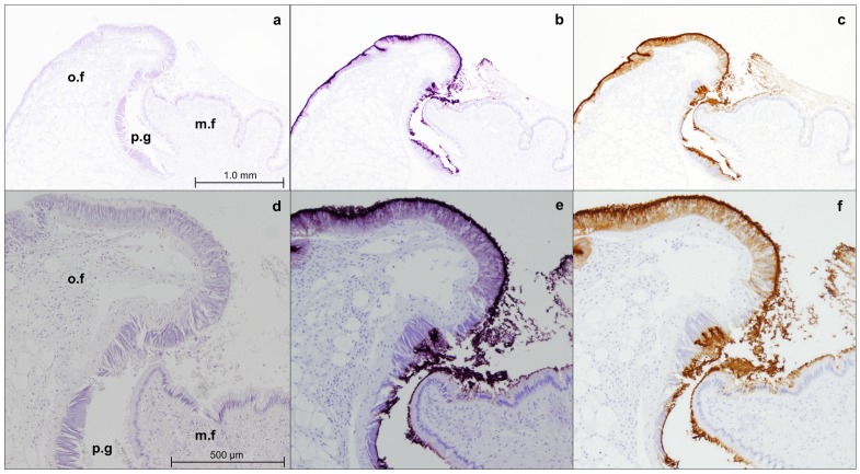
Fig 3. Immunostaining of the mantle edge of adult M. galloprovincialis with M22.8 mAb.
Microphotographs were taken at 4× (a, b, c) and 10× (d,e,f). (o.f): outer fold; (m.f): middle fold; (p.g): periostracal groove; (a,d) negative control; (b,e): immunostaining revealed with VectorVip; (c,f) immunostaining revealed with DAB.

