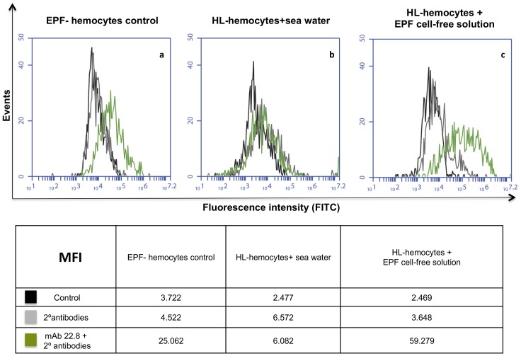
Fig 8. Flow cytometry analysis of permeabilized hemocytes.
(a) EP hemocytes used as positive control; (b) hemolymph hemocytes exposed to sea water; (c) hemolymph hemocytes exposed to EPF cell-free solution. Cells were incubated with the M22.8 mAb followed by FITC-labelled secondary antibodies. (MFI): median fluorescence intensity; (HL): hemolymph.

