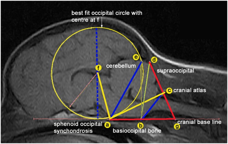
Fig 1. Midline sagittal T1-weighted MRI of brain and craniocervical region of a female GB backcross.
A framework of measured lines and angles is used to assess conformational features associated with CM verified in a previous study [1]. (A) dorsum of spheno-occipital synchondrosis (B) basion of basioccipital bone (C) rostral edge of the dorsal lamina of the atlas (D) junction between the supraoccipital bone and the occipital crest (E) most dorsal point of intersection of the cerebellum with the occipital lobe circle (F) centre of occipital lobe circle placed on the extended cranial baseline (AB) (G) intersection point with the extended AB baseline and DC. The five traits measured in the study are lines ae, bc and f-diameter (blue) and angles FAC (yellow) and AGD (red).

