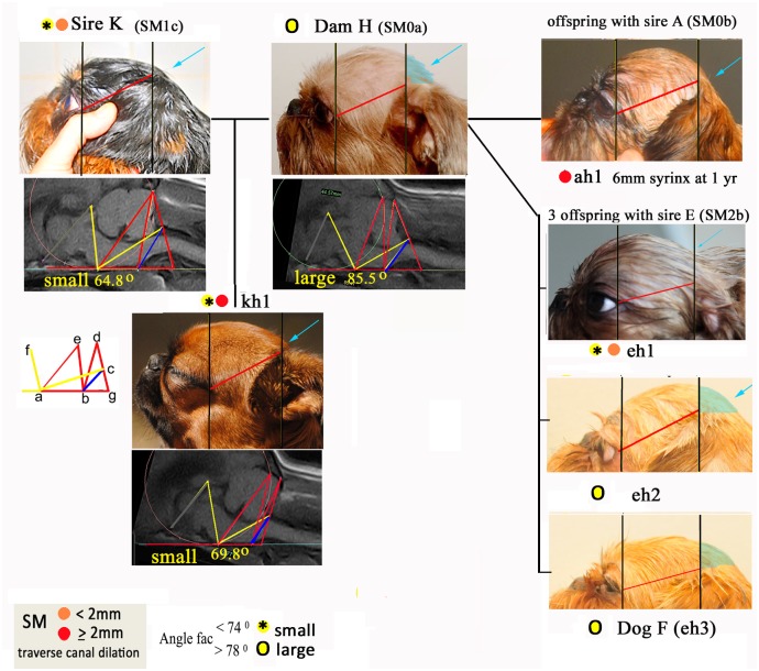
Fig 5. Head conformation and associated angle FAC in six relatives of Bitch H with and without SM.
TW1 sagittal MRI of the caudal fossa and cranial-cervical junctions with superimposed morphometric framework of lines and angles for parents and offspring enhances comparison. The differences in the size of angle FAC are reflected in the lack of skull development caudal to the ear pinna (behind the ears) for dogs with SM compared to dogs with no SM (‘normal’ caudal skull shaded aqua colour). The photos of the heads have been resized to allow comparison using two vertical lines (black) placed at the outer eye and the origin of the external pinna (red) a consistent distance apart.

