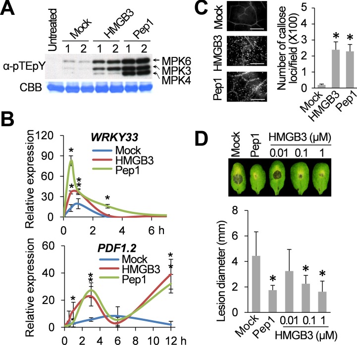
Fig 1. Extracellular HMGB3 activates innate immune responses.
A. HMGB3- or Pep1-induced MAPK activation in Arabidopsis. Leaves were collected 15 min after infiltration with water containing either 1 μM recombinant HMGB3 (HMGB3) or Pep1 peptide (Pep1) for the MAPK activation assay. Activation of MPK3, MPK4, and MPK6 by MAPK kinase-mediated phosphorylation of the TEY sequence was detected with α-pTEpY antibody using immune-blot (IB) analyses. Ribulose-1,5-bisphosphate carboxylase/oxygenase (Rubisco) large subunit protein stained with Coomassie Brilliant Blue (CBB) served as a loading control. 1 and 2 denote independent biological replicates. B. RT-PCR analysis of WRKY33 (upper panel) and PDF1.2 (lower panel) expression at the indicated times after infiltration with water containing either 0.1 μM HMGB3 or Pep1. Expression levels were plotted relative to the expression in untreated leaves. Data are the mean ± SD (n = 4). C. HMGB3- or Pep1-induced callose deposition in Arabidopsis. Leaves were stained with aniline blue 15 h after infiltration with water containing either 0.1 μM HMGB3 or Pep1. Representative pictures are shown in the left panel. Bars = 100 μm. Data are the mean ± SD (right panel, n = 20). D. HMGB3- or Pep1-induced resistance to B. cinerea. Leaves were infiltrated with water containing the indicated concentrations of HMGB3 or 1 μM Pep1 one day before B. cinerea inoculation. Representative disease symptoms at 3 days post infection (dpi) are shown in the upper panel. Data corresponding to this time point are presented as the mean ± SD (lower panel, n = 6). Leaves infiltrated with water served as mock control in all experiments. Asterisks in B, C, and D indicate significant differences from the mock-treated leaves (t test, P < 0.05).

