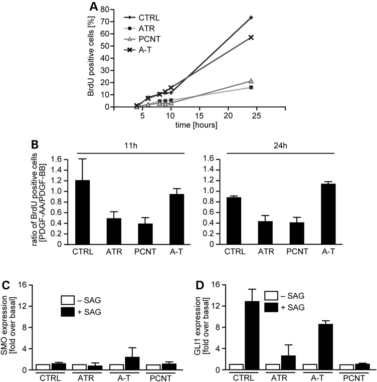
Figure 2.
ATR-SS cells show impaired cilia-dependent growth-factor signalling. (A) Patient-derived hTERT fibroblasts were serum-depleted for 48 h. Following serum and BrdU addition, the percentage of BrdU+ S phase cells was monitored as indicated. At least 200 cells were monitored. (B) The indicated hTERT fibroblasts were serum-depleted for 48 h. PDGF-AA or -BB and BrdU were then added and the percentage of BrdU+ cells (i.e. cells that have entered S phase) estimated by IF 11 and 24 h later. Results are expressed as the ratio of BrdU+ cells following treatment with PDGF-AA versus PDGF-BB. A ratio of 1 demonstrates equal signalling from PDGF ligand isoforms (-AA or -BB) and <1 demonstrates an impaired response to PDGF-AA. The percentage of BrdU+ cells after PDGF-BB treatment after all siRNA treatments is shown in Supplementary Material, Figure S1. This verifies that the indicated siRNA does not impact upon PDGF-BB signalling. Results represent the mean ± SD of three experiments. (C and D) qRT–PCR analysis of SMO and GLI1 transcript levels in the indicated cells with or without SAG at 24 h. SMO is a reference as it does not change after SAG addition. The WT and PCNT data are part of a previously published data set (16). The analysis of ATR- and A-T-deficient cells was carried in the same experiments and has not been previously published. Results represent the mean ± SD of three experiments.

