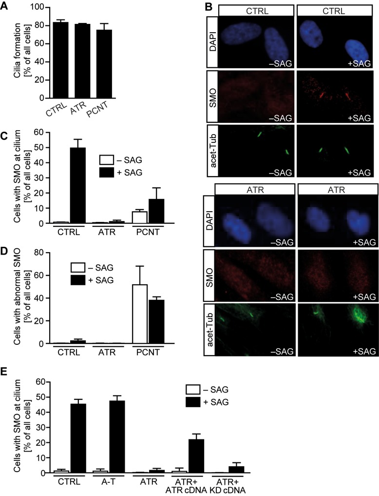
Figure 6.
ATR-SS cells show defects in SMO recruitment to cilia and GLI1 transcript activation. Patient-derived hTERT fibroblasts were serum-depleted for 48 h. SAG was then added for 24 h. (A) Under this prolonged condition of serum starvation (72 h), cilia form normally in all cell lines. (B) In control cells, in the absence of SAG, SMO localized diffusely and not specifically at cilia. In the presence of SAG, strong uniform SMO staining is observed along the cilia length (detected using acetylated tubulin). (C) The percentage of cells with co-localized cilia and SMO with or without SAG treatment. (D) The percentage of ciliated cells with abnormally localized SMO with or without SAG treatment. (E) SMO localization at cilia in ATR-SS cells can be restored following expression of ATR cDNA but not kinase dead ATR cDNA (KD). Results represent the mean ± SD of three experiments. WT and PCNT data are part of the data set previously published in Stiff et al. (16).

