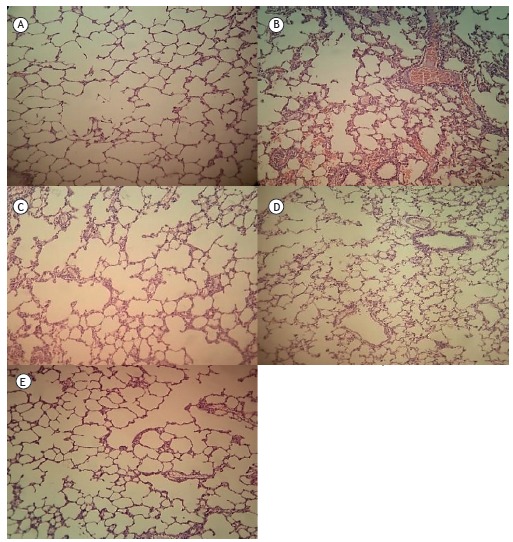
Figure 5. Lung tissue samples stained with hematoxylin and eosin (original magnification, ×100): (A) control group sample, showing no remarkable pathological changes; (B) ischemia/reperfusion (I/R) group sample, showing widespread histological changes such as edema, severe alveolar congestion, alveolar collapse, and inflammatory cell infiltration; and (C, D, and E, respectively) I/R+N-acetylcysteine, I/R+pentoxifylline, and I/R+N-acetylcysteine+pentoxifylline group samples, all showing fewer histological alterations (markedly less interstitial edema and inflammatory cell infiltration) in comparison with the I/R group sample.

