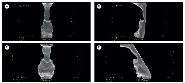
Figure 3. Cervical CT for determining upper airway volume-in the neutral position (without head elevation): anterior view (in A) and lateral view (in B); and with the head elevated by 44° with a wedge: anterior view (in C) and lateral view (in D). Volume was measured from the hard palate to the base of the epiglottis.

