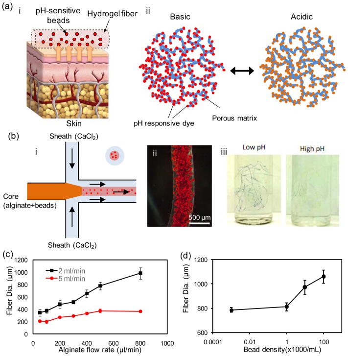
Figure 1.
Fabrication of pH-sensing microfibers. (a) A schematic illustration of the pH-sensing hydrogel microfiber designed for long-term epidermal monitoring (i) and the action mechanism of the mesoporous silica particles containing pH-responsive dye with electrostatic interaction to the solid matrix of the mesoporous particles (ii). (b) Schematic of the fiber fabrication process using coaxial streams of Na-alginate mixed with dye loaded beads surrounded with CaCl2 solution (i). A typical bead-laden alginate fiber fabricated using the microfluidic chip (ii). A representative micrograph of pH-responsive bead-laden hydrogel microfibers (iii). (c) Effect of core flow rate on the fiber diameter at two CaCl2 flow rates of 2 and 5 mL/min. (d) Effect of the bead density on fiber diameter; the core and sheath flow rates were constant at 600 μL/min and 2 mL/min, respectively.

