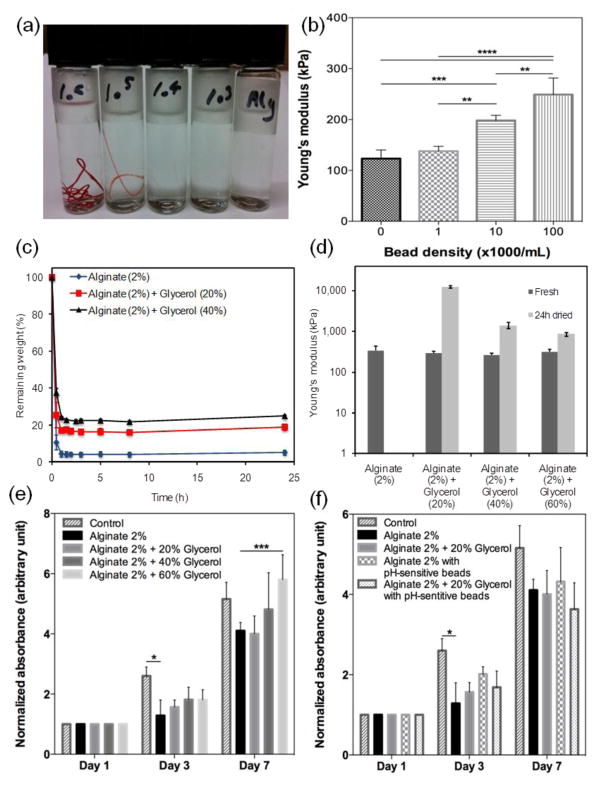
Figure 2.
Physical characterization of the pH-sensing fibers. (a) Visual appearance of fibers with various bead densities; from right to left: 0, 1×10³, 1×10⁴, 1×10⁵, 1×10⁶. (b) Variation of fiber mechanical strength as a function of bead concentration. (c) Effect of glycerol concentration on the dehydration rate of hydrogels. (d) Variation of fiber mechanical properties immediately after fabrication and after 24 hr dehydration at 37 °C. Metabolic activity of cells cultured with hydrogels containing various (e) glycerol and (f) bead concentrations.

