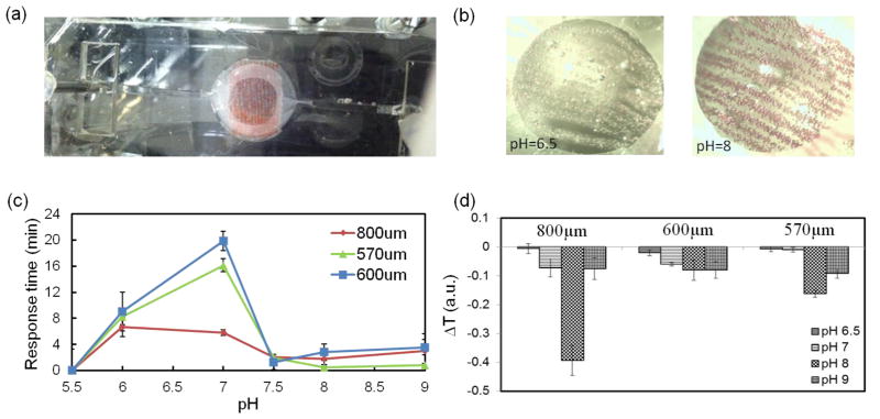
Figure 3.
UV-VIS-NIR USB spectrometer data of the response of the fabricated fiber to pH variation. (a) array of aligned fibers composed of brilliant yellow doped microbeads were placed in a polydimethylsiloxane (PDMS) chamber and exposed to different pH environments. (b) different colors of the pH-responsive fibers at solutions with pH values of 6.5 (yellow) and 8 (red). (c, d) Variation of the response time and detection signal as a function of fiber diameter and different pH environments, respectively.

