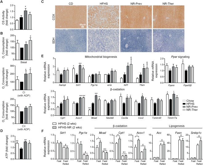
Figure 4.

Treatment with NR increases liver tissue respiratory capacity ex vivo and β‐oxidation gene expression. Measurements were performed on liver samples from CD, HFHS, NR‐Prev, and NR‐Ther cohorts after 9‐18 weeks of treatment (NR dose: 400 mg/kg/day) and are represented as fold changes compared to the HFHS group. NR increased (A) citrate synthase activity, (B) basal OCR and CI‐coupled driven OCR, in the presence of glutamate, pyruvate, malate, and ADP, but was not significant for CII‐coupled driven OCR, in the presence of succinate, although a trend was noted (n = 6). (C) Histological sections show improvements in both COX and SDH activities in NR‐treated groups compared to HFHS‐fed mice (n = 4), culminating in the attenuation of the decline in (D) ATP levels (n = 5). (E) Increases were observed for mRNA transcript levels of genes regulating mitochondrial biogenesis, Pparα and Pparδ/β, and genes regulating β‐oxidation (n = 6). (F) Liver gene transcript expression measurements were performed for Sirt1, Pgc‐1, and genes associated with β‐oxidation and lipogenesis in mice given 2 weeks of an HFHS or HFHS‐NR diet (NR dose: 400 mg/kg/day). Starting at 8 am, mice were fasted for 24 hours or fasted for 18 hours and refed for 6 hours. Data are represented as fold changes compared to the fasted CD group. For panels A, B, D and E: *P < 0.05; **P < 0.001; ***P < 0.0001 compared to the HFHS cohort and ϵ P < 0.05 compared to the CD cohort. For panel F: ϵ P < 0.05, overall effect of treatment versus control mice; ‡P < 0.05, interaction of each treatment versus control mice. Data in all panels are expressed as mean ± SEM. One‐way ANOVA with a post‐hoc Bonferroni test was used for statistical analyses of panels A, B, D, and E. Two‐way ANOVA with a post‐hoc Holm‐Sidak test was used for statistical analysis of panel F. Male mice were used for these experiments.
