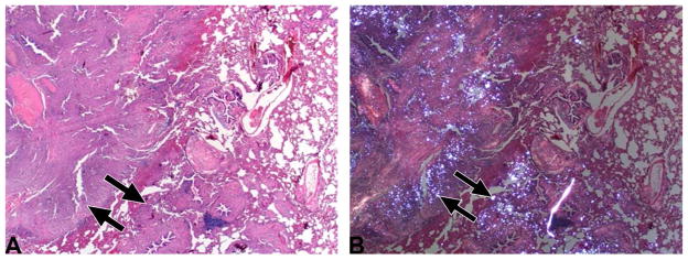
Figure 4. Pathological confirmation of silica deposits and disease development.
(A) Histopathological appearance of the lung (from Subject 4, see also Figure 1D) showed locally extensive remodeling accentuated by fibrosis (arrows) and inflammation (HE stain, 20x). (B) Same section as Figure 4A, but examined with polarized light. Note that the white (refractile) silica granules (arrows) are admixed within pulmonary remodeling (HE stain, 20x).

