Figure 2.
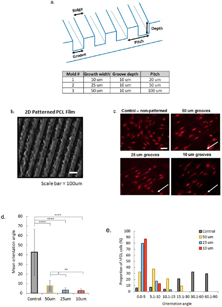
Micropatterned 2D PCL film design was used to determine effects of groove width on hPDL cell alignment in vitro. Films were patterned using three different molds (a) to embed grooved features onto the polymer surface (b, surface with 50um wide grooves, Nikon SMZ18 stereo microscope). DilC(12)-3 stained hPDL cells seeded on hydrolyzed and fibronectin treated PCL films with non-grooved (control) and grooved surfaces (c). White arrows indicate direction of grooves (scale bar=50um). Average orientation of hPDL cells on control (non-patterned), 50 um, 25 um, and 10 um grooved PCL films, where 0° indicates complete alignment with pattern and 90° indicates a cell that is perpendicular to a groove (d). Proportion of hPDL cells within a specific orientation angle for all PCL film groups, with 0–5° groups indicating highest alignment of cells within grooved microfeatures (e). (Error bars: ± SD; * p<0.05; ** p<0.01; **** p<0.0001).

