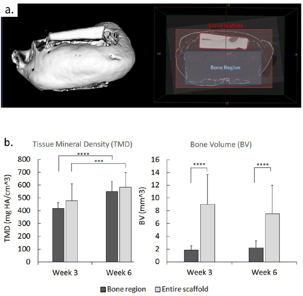
Figure 4.
Formation of bone in the 3D printed scaffold region seeded with adenoviral BMP7 transduced gingival fibroblasts. Micro-computed tomography (uCT) of implanted scaffolds was used for bone volume (BV) and tissue mineral density (TMD) analysis of osseous tissue growth in vivo in the bone region of the scaffold (a) compared to the formation of tissue in the entire scaffold external to bone region at 3 and 6 weeks post-harvesting (b). (Error bars: ± SD; *** p<0.001, **** p<0.0001; n=5–6).

