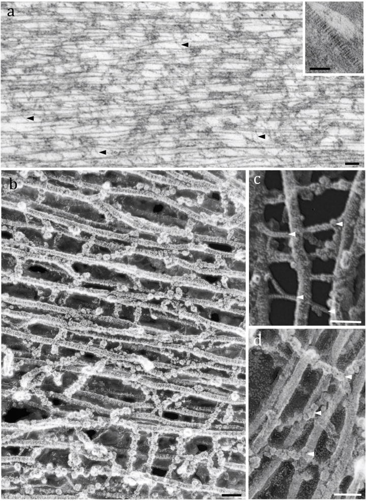
Figure 2.
Fine structure of the TM striated sheet: Fig. 2a: TEM of a rat TM showing the parallel-arranged type II collagen fibrils interwoven by thin filaments (arrowheads). Inset: TEM of an individual collagen fibril with its typical banding pattern. Fig. 2b: High-resolution freeze-etching image of a guinea pig TM formed by collagen fibrils crosslinked by numerous filamentous materials. Fig. 2c: Detail image of collagen fibrils crosslinked by thin filaments (arrowheads). Fig. 2d: High magnification showing collagen fibrils connected by globular-arranged filaments. Scale bars: a = 100 nm; Inset, b, c and d = 50 nm

