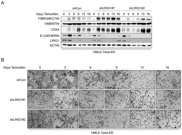
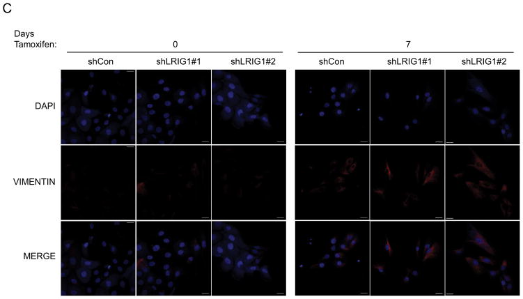
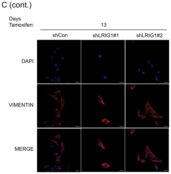
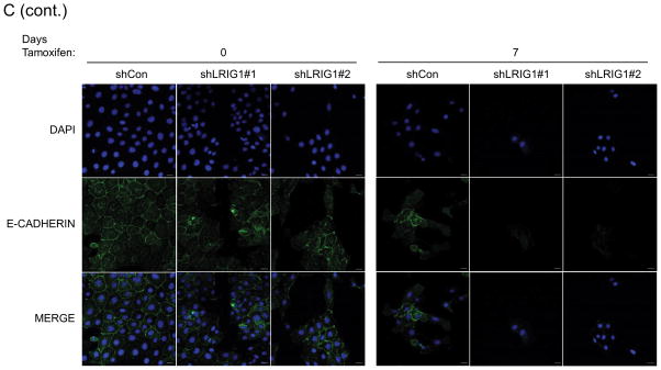
Figure 3. LRIG1 knockdown accelerates EMT of human mammary epithelial cells.
(A) Western blot analysis of total cell lysates collected from stable pooled clones of HMLE-Twist-ER cells expressing control shRNA (shCon) or LRIG1-targeted shRNAs (shLRIG1# 1 and shLRIG1#2). Cell lysates were prepared pre-Twist induction (Day 0) and post-Twist induction (Days 3, 6, 9, 13, 16). Lysates were blotted as indicated with Actin as a loading control. (B) Images (10x objective) of HMLE-Twist-ER-shCon and HMLE-Twist-ER-shLRIG1#1 and shLRIG1#2 cells pre-Twist induction (Day 0) and post-Twist induction (Days 3, 6, 9, 13, 16). Scale bar = 20 μm. (C) Confocal immunofluorescence analysis of HMLE-Twist-ER-shCon and HMLE-Twist-ER-shLRIG1#1 and shLRIG1#2 cells pre-Twist induction (Day 0) and post-Twist induction (Days 7 and 13). Staining for Vimentin and E-cadherin, as shown. All data are representative of at least 3 independent experiments. Scale bar = 20 μm.


