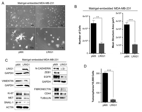
Figure 8. LRIG1 suppresses invasive 3D morphology of MDA-MB-231 breast cancer cells.
(A) Phase-contrast images (4x and 10x objectives, as indicated) of MDA-MB-231-pMX control (left) and MDA-MB-231-pMX-LRIG1 (right) cells grown in 3D Matrigel culture for 7 days. (B) Left panel: Proliferation of MDA-MB-231-pMX control and MDA-MB-231-pMX-LRIG1 cells was measured by manually counting cells recovered from 3D Matrigel culture after 7 days. Right panel: The area of structures observed in (A) were quantified (n ≥ 80 structures). (C) Western blot analysis of total cell lysates from MDA-MB-231-pMX control and –LRIG1 cells recovered after growing in 3D Matrigel culture for 7 days. (D) Quantification of tumorspheres formed by MDA-MB-231-pMX control and MDA-MB-231-pMX-LRIG1 cells. Data are presented as mean ± SEM, collected from at least 3 independent experiments. (** = p<0.01–0.001, *** = p<0.001 by Student’s t-test).

