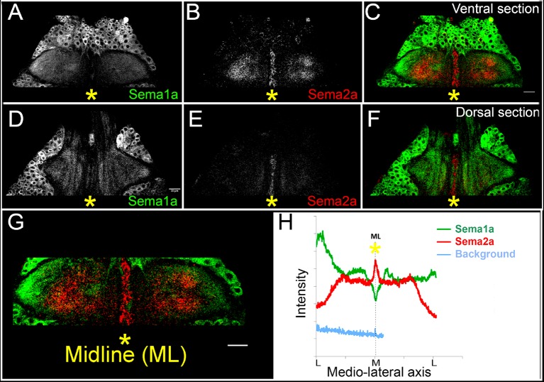
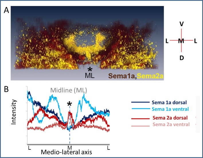
Figure 5. Sema2a is highly expressed in the ganglionic midline and intermediate region.
Immunocytochemical analysis of expression of Sema1a and Sema2a in prothoracic ganglion at 25h APF (after puparium formation). (A–C) Ventral section. (D–F) Dorsal section. (A, D) Sema1a immunolabeling. (B, E) Sema2a immunolabeling. (C, F) Overlay of Sema1a and Sema2a immunolabeling. (G) Magnified view of neuropile. (H) Intensity profile of Sema1a and Sema2a immunolabeling taken along mediolateral axis of neuropile (in Z-stack; overlay of all optical sections). Yellow asterisk is placed at the ganglionic midline (ML). Sema2a level is highest at the midline and high in the intermediate regions in the neuropile and very low at the lateral edge. Scale bar = 20 microns.


