. 2016 Feb 25;5:e12204. doi: 10.7554/eLife.12204

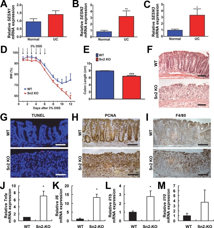
Figure 1. Protective function of Sestrin2 against colon injury.

(A-C) Upregulation of human SESN2 and SESN3 expression in ulcerative colitis (UC). mRNA expression of human SESN1-3 was analyzed through quantitative RT-PCR of non-inflamed (Normal) and inflamed (UC) colon tissues from patients with UC (n=10; mean ± s.e.m.). These samples were histologically confirmed and formerly described (Xue et al, 2013). (D-M) Loss of Sestrin2 impairs recovery from DSS-induced colitis in mice. 6-month-old WT and Sesn2-/- mice (n=4 each) were treated with 3% DSS in drinking water for 6 days (arrows), followed by 6 days of regular water. Body weight was measured over 12 days (D; mean ± s.e.m.). At the final day of the experiment, mice were sacrificed and colon length was measured (E). The data are shown as the mean ± s.e.m. The colons were isolated and fixed for H&E staining (F), TUNEL staining (G), PCNA staining (H) and F4/80 staining (I). The levels of the indicated mRNAs, which are indicative of active inflammation, were quantified by real-time PCR (J-M; mean ± s.e.m.). *p<0.05, **p<0.01, ***p<0.001. P values are from Student’s t-test. Scale bars, 100 μm.

DOI: http://dx.doi.org/10.7554/eLife.12204.003

Figure 1.

Figure 1—figure supplement 1. Hypersensitivity of Sesn2-/-/Sesn3-/- mice against DSS-induced colon injury.

Figure 1—figure supplement 1.

1-year-old WT and Sesn2-/-/Sesn3-/- mice (n=4 each) were treated with 3% DSS in drinking water for 7 days (arrows in A), followed by 5 days of regular water.Body weight was measured over 12 days (A). At the final day of the experiment, mice were sacrificed and colon length was measured (B). The colons were isolated and fixed for H&E staining (C), TUNEL staining (D), PCNA staining (E) and F4/80 staining (F). Data are shown as the mean ± s.e.m. **p<0.01. P values are from Student’s t-test. Scale bars, 100 μm.
Figure 1—figure supplement 2. Acute colon injury is comparable between WT and Sesn2-/- mice during DSS treatment.

Figure 1—figure supplement 2.

2-month-old WT and Sesn2-/- mice (n=4 each) were treated with 3% DSS in drinking water for 7 days (DSS – 7d only).At this time point, mice were sacrificed and colon length was measured (A). The data are shown as the mean ± s.e.m. The colons were isolated and fixed for H&E staining (B). The levels of Tnfa and Xbp1s mRNAs, which are respectively indicative of active inflammation and ER stress, were quantified by real-time PCR (C,D; mean ± s.e.m.). Scale bars, 200 μm.