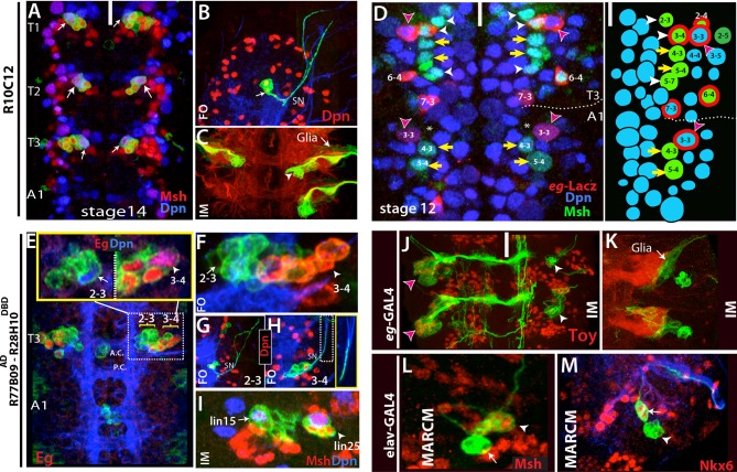
Figure 5. Thoracic-specific NB2-3 and NB3-4 generate lineage 15 and lineage 25, respectively.
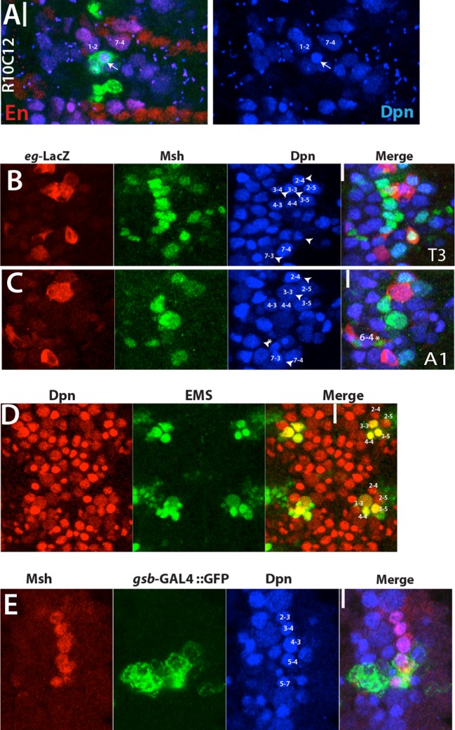
(A) R10C12 marks uniquely Msh+ NB2-3 (arrows) in each thoracic hemisegment but not in abdominal ganglia. Dpn, blue; Msh, red (B) A lineage clone containing NB2-3 (arrow) extends only efferent axons, which splits into two bundles after exiting the CNS via the segmental nerve (SN). (C) Immortalization of R10C12 expression in the NB2-3 progeny marks lineage 15, which is composed of motor neurons (arrowhead) and glia (arrow). (D) T3 and A1 segments from a stage 12 embryo shown. eg-lacZ (red) and Msh (green) co-staining reveal differences in NB (blue) pattern of T3 and A1 segments. Schematic representation is shown on the right. Msh marks three thoracic specific NBs (NB2-3, NB3-4, and NB5-7; white arrowheads). NB3-4, which is marked by both eg-lacZ and Msh, resides medial to NB3-3. In abdominal segments, no NB with lacZ and Msh expression is detected medial to NB3-3; note the region immediately medial to NB3-3 does not have any NB (asterisks). (E-I) R77B09AD-R28H10DBD used to identify the postembryonic progeny of NB2-3 and NB3-4. (E-G) Nerve cords from late stage embryos shown. NB3-4 (arrowhead) and its progeny express Eg (red) while NB2-3 (arrow) and its progeny do not. Inset shows the magnified views of NB2-3 (left) and NB3-4 (right) lineages. (F) A lineage clone via R77B09AD-R28H10DBD separates two NB lineages: NB3-4 lineage is marked with red. Progeny of NB3-4 (H) extend axons out of the CNS using the SN route like NB2-3 progeny (G) but do not extend them as far as NB2-3 progeny (see inset H). (I) Nerve cord from a late-stage larva shown. Msh, red; Dpn, blue. Immortalization of R77B09AD-R28H10DBD expression marks lineage 15 (NB2-3 progeny) and lineage 25 (NB3-4 progeny). (J) Immortalization of eg-GAL4 marks Toy expressing lineage 8 (NB3-3 progeny; red arrowheads; see also Figure 4—figure supplement 1L) and lineage 25 (white arrowheads). (K) Lineage 25 contains glial cells (arrow). (L-M) elav-MARCM clones showing lineage 25 have a small cluster of Msh expressing cells (arrowheads) and two Nkx6 expressing motor neurons (arrows). Msh expressing cells reside closely to Msh+ NB3-4, but do not extend any processes (arrowheads in L and M). Note glial cells in lineage 25 are not visible in MARCM clones since elav-GAL4 does not mark glial cells. White bar indicates the midline. FO, flip-out lineage clone; IM, immortalization; A.C., anterior commissure; P.C., posterior commissure; white bar, midline.


