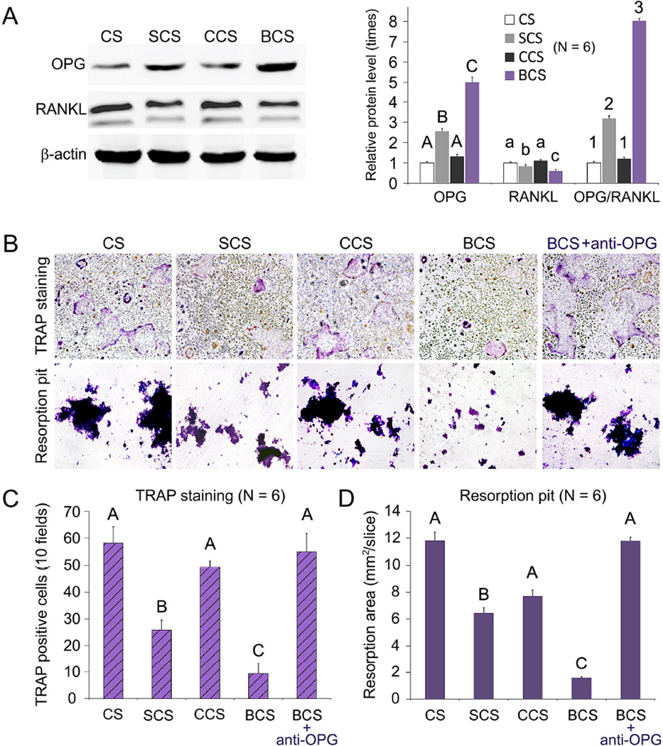
Figure 4.
Effects of scaffold-conditioned osteoblasts on RANKL-mediated osteoclastogenesis by RAW 267.4 cells. A. Western blot analysis (left) of OPG and RANKL secreted by mMSCs exposed to different scaffolds (CS, SCS, CCS or BCS). Expression levels of OPG, RANKL and OPG/RANKL ratio of the different groups were normalized against β–actin. For each parameter, groups identified with the same upper case letters, lower case letters or numbers are not significantly different (P > 0.05). B. Representative images of TRAP-stained osteoclasts and resorption pits produced by RAW 267.4 cells co-cultured with scaffold-conditioned osteoblasts in the presence of RANKL (50 ng/mL; magnification 40×). The inhibition effect produced by anti-OPG antibody is shown on the far-right. C and D. Bar charts depicting the number of TRAP-positive multinucleated cells (10 fields) and area of resorption pits. For each chart, groups identified with the same upper case letter are not significantly different (P > 0.05).

