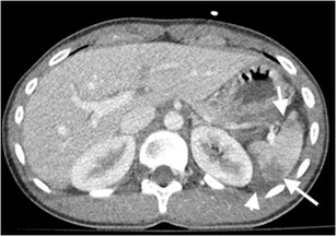
Fig. 1.

Splenic hematoma. Axial contrast-enhanced CT of a 35-year-old man after a motor vehicle accident (MVA) demonstrates hematomas in splenic parenchyma with low-attenuated appearance (arrows). Perisplenic haemorrhage is also noted (arrowhead). The splenic capsule is intact
