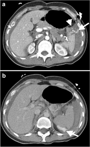
Fig. 3.

Active bleeding after splenic injury. Axial contrast-enhanced CT images at early (a) and delayed (b) phase demonstrate splenic laceration (arrowhead in a) and active intraperitoneal bleeding (long arrows in a and b). Absence of clot formation results from active massive haemorrhage, as clot formation is not fast as bleeding. Extravasated contrast material can accumulate in a dependent part of the body without restriction of any clotted hematoma as of yet (arrow in b). Moreover, the presence of perihepatic fluid (short arrow in a ) supports the evidence of massive bleeding, since blood can act in a pattern similar to the free fluid, due to a decreased percentage of clot formation
