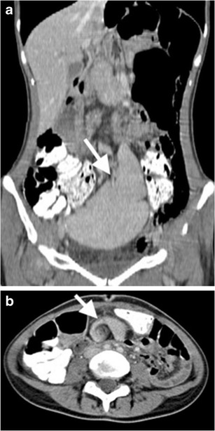
Fig. 9.

Splenic torsion in a patient with wandering spleen. (a) Coronal contrast-enhanced CT image reveals ‘ectopic wandering spleen’ (arrow) in the pelvis. (b) Axial contrast-enhanced CT at superior level than pelvis further demonstrates ‘whirling’ appearance (arrow) of splenic vessels, suggesting ‘torsion of the spleen’ as well. Normal parenchymal enhancement indicates lack of infarction
