
Fig. 6.
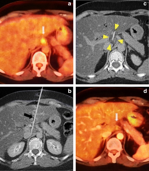
Cryoablation of a peripancreatic lymph node. a In a 44-year-old patient after surgical treatment of ovarian carcinoma, PET-CT image shows a lymph node metastasis (white arrow) near the pancreas. Since the metastasis was solitary, percutaneous ablation was planned, and because of the critical location of the lesion, cryoablation was preferred. b Under local anaesthesia and US+CT guidance, a cryoablation needle (black arrow) was inserted transhepatically into the lesion. c The lymph node was ablated with two freeze and one thaw cycle, and an iceball was typically seen (arrowheads). Since the liver was traversed, track ablation was performed after cryoablation to prevent bleeding and tumour seeding. d Eight months later, the follow-up CT shows complete metabolic response of the lesion (white arrow)