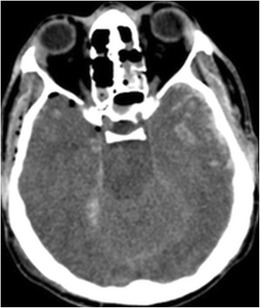
Fig. 14.

A 19-year-old male, following a high energy motor vehicle accident with hemorrhagic contusions in the temporal lobes bilaterally. Note pneumocephalus anterior to the right temporal lobe, small amount of subarachnoid blood along the right tentorial leaf and subarachnoid blood along the left temporal lobe. Additionally, there is relative sulcal effacement and loss of grey-white matter differentiation from diffuse brain swelling
