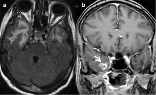
Fig. 15.

A 42-year-old female with a history of radiation for nasopharyngeal carcinoma. Axial FLAIR image (a) shows vasogenic edema in the anterior aspect of the temporal lobes bilaterally, right greater than left. Coronal contrasted T1 weighted image (b) shows enhancement on the right (arrow). On biopsy, the diagnosis of radiation necrosis was confirmed
