
Fig. 16.
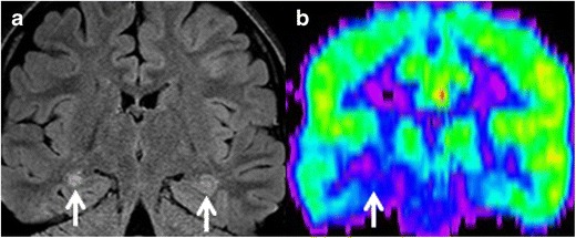
(a) Coronal FLAIR image of a 31-year-old man with partial complex seizures showing bilateral FLAIR hyperintensities in hippocampal body, right greater than left (arrows), suspected for bilateral mesial sclerosis. PET study (b), show unilateral hypometabolic focus (arrow), compatible with right-sided mesial temporal sclerosis